
2.25million/year
Estimated annual volume of core microsurgical procedures in China: approximately 1.1545 million neurovascular, 512,100 malignant brain tumor related, 300,600 ENT, and 283,800 ophthalmic (excluding cataract).
SPECTRA NAVIGATION
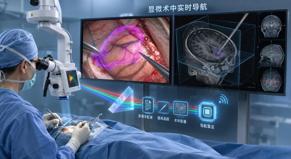
Microsurgical navigation for neurosurgery and microsurgery. It mounts onto the microscope already in the room, combining split-path stereo imaging, micron-scale instrument tracking and dynamic registration.
Navigation has to keep answering “where am I”. Existing systems register once, before surgery, and fail the moment tissue deforms — continuous intraoperative registration remains unsolved.
The project remains in R&D and validation · Core technologies validated on the bench and in animal studies; end-to-end system navigation accuracy will be established by third-party testing after integrated prototype V1
PROJECT FILM
A full walkthrough of the operating principle, the prototype, and the experimental setup (with narration).
Clinical Need
The relative positions of lesion boundaries, instrument tips, and critical anatomy are difficult to judge directly. Critical boundaries and residual tissue are often below 0.5 mm — beyond the resolving limit of the human eye and the depth of field of an operating microscope — while conventional navigation accuracy of 1–3 mm sits orders of magnitude away from the 20–25 µm scale of microsurgery. As procedures move deeper and toward smaller structures, reliance on individual experience increases, along with training time and variability in risk.
Estimated annual volume of core microsurgical procedures in China: approximately 1.1545 million neurovascular, 512,100 malignant brain tumor related, 300,600 ENT, and 283,800 ophthalmic (excluding cataract).
Reported mean survival benefit of gross total versus subtotal resection for grade IV glioma.
Density is about 30% of that in developed countries, with a 10–15 year training cycle. In microvascular free-flap surgery, for example, novice anastomosis patency is only 60–70%, and roughly 600 cases are needed before the success-rate curve plateaus. Navigation supports decision-making and shortens the learning curve.
Procedure-volume data: China Health Statistics Yearbook. Other clinical figures are drawn from public research; definitions, reference years, and indication scope may vary between sources.
Limits of Existing Solutions
Conventional navigation is designed around rigid bony fixation, large reflective markers, and an open field. All three premises break down in the microscopic field — drift, scale, and features are the three limits that follow, and the left column of the next section, Product Concept, is where they are drawn together.
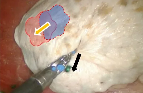
CSF release and resection continuously shift tissue position. Existing navigation cannot re-register in real time, so the on-screen position gradually diverges from the actual surgical field.

Existing navigation carries system error of roughly 1–3 mm, insufficient for microscale boundary judgment. Conventional reflective spheres are about 11 mm in diameter against 0.3 mm microsurgical instrument tips, and readily obstruct a narrow field.

A microscopic field spans only a few millimeters and offers very few stable, repeatable features. Glare, bleeding, occlusion, and soft-tissue deformation further alter the local image.
Product Concept
Navigation rests on two conditions: where the instrument is right now and what the tissue in front of it is. Both must hold throughout resection. A system that answers them once, before the incision, is providing a plan — not navigation.
Objective: bring boundary, distance, and risk cues into the current view without disrupting the surgeon's core operating habits.Product value still requires continued validation in real clinical workflows, against defined baselines, and through surgeon feedback.
Core Technology · Product System
Visualization reconstructs the 3D surgical field, precision tracking localizes the instrument tip, and intelligent registration updates the coordinate reference as tissue and the field change. These capabilities connect along a single navigation chain, each embodied in one component of the system.
Product System
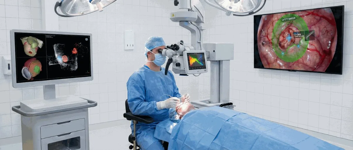
The system mounts onto the operating microscope already in the room and changes neither staff positions nor equipment layout. The three components marked here, plus the overlay inside the surgical field, are all of it that is visible in the operating room.
Select a numbered point in the scene, or any component below, to mark its position and open the technology behind it, with its study records.
01 · VISUALIZATION

One optical path, two purposes. The three images show how visualization is implemented: coaxial split-path visible and near-infrared acquisition, a validation prototype that establishes 3D metrology, and final integration with an operating microscope. The split-path camera module has completed bench validation.


The microscope platform combines imaging and metrology, preserving the live surgical field while providing structural, depth, and relative-position references.
02 · PRECISION
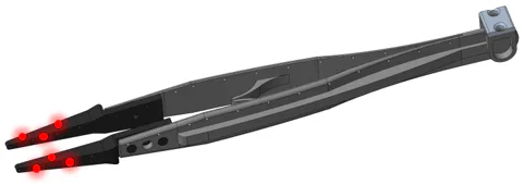
Ultrafine optical fibers are embedded in microsurgical forceps, scissors, and related instruments to form active tracking points while preserving the original dimensions, weight, and handling. Tracking remains available in deep and narrow fields, and tip position is linked to the preoperative model.
03 · INTELLIGENCE

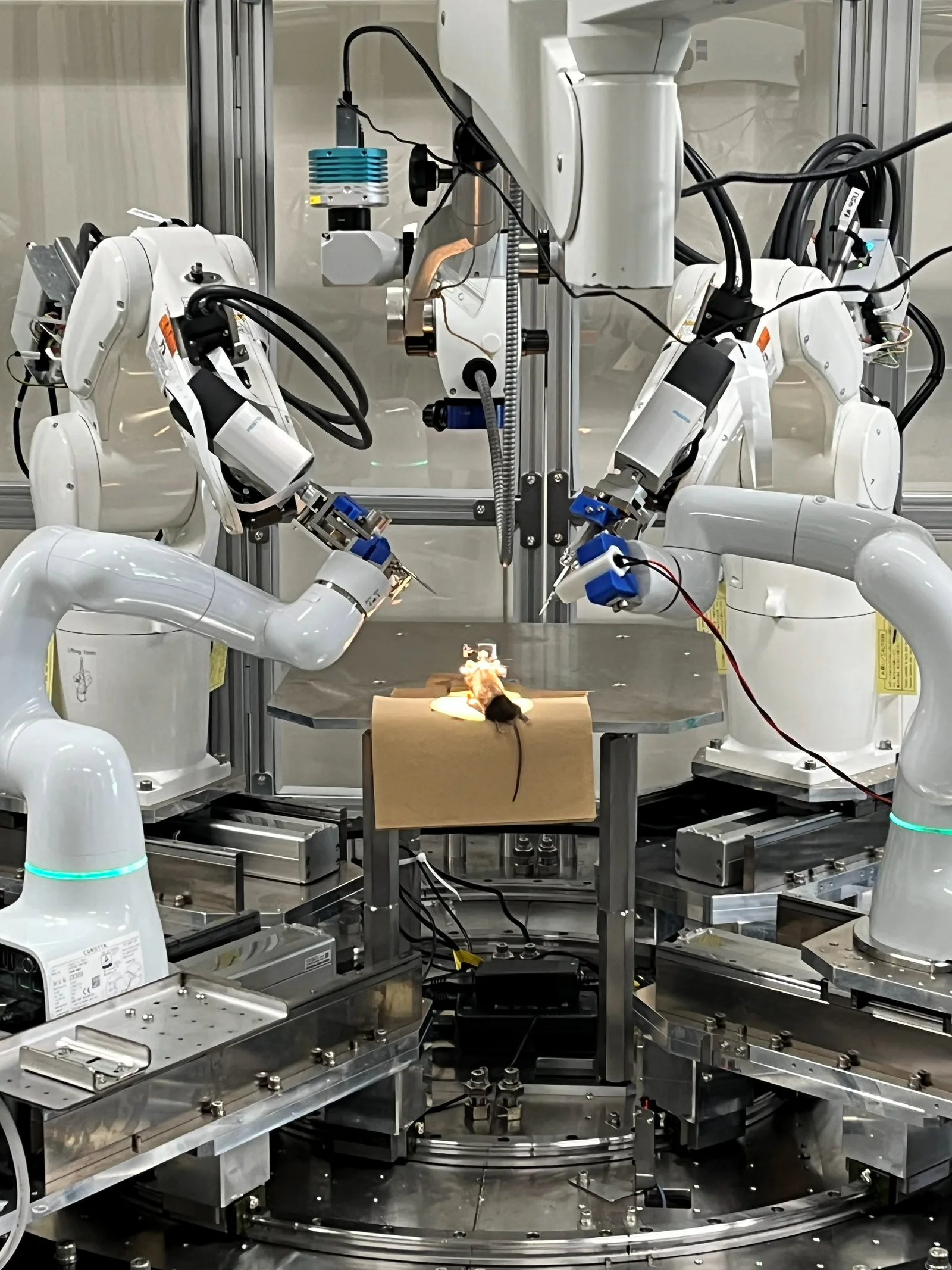
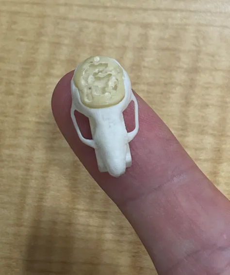
Vessel orientation and bone-surface texture serve as semantic alignment constraints, combined with a dynamic-landmark mechanism that continuously assesses confidence and updates the reference online. Core algorithm accuracy has been validated on a robotic platform and across 74 mouse craniotomy experiments (ethics approval A2023M042-07, the University of Tokyo).
Extract constraints from recognizable structures such as vessels and bone-surface texture to align the preoperative model with the intraoperative view.
Under a small field, occlusion, glare, and tissue change, the system continuously checks whether the current reference remains reliable.
Dynamic landmarks update relative positions and return the instrument trajectory, model, and live image to a common spatial relationship.
BENCHMARK · REGISTRATION ERROR
Under microscopic deformation, the errors reported for existing registration methods differ from our measured values by an order of magnitude. The table places the three approaches side by side and labels the source of each figure.
| Registration method | Reported error | Microscopic deformation | Source |
|---|---|---|---|
| Bone-anchored registration | approx. 0.8–2 mm | Not applicable | Published literature |
| Conventional ICP surface matching | 2.7–5.3 mm | Not applicable | Published literature |
| This project · dynamic registration | 0.164 ± 0.059 mm (max 0.270 mm) | Applicable | Measured in mouse studies |
Literature values and our measured values come from different experimental conditions, sample sizes, and statistical definitions, and are not directly equivalent; the comparison is shown to indicate the difference in error magnitude. Project data come from mouse craniotomy studies; the full statistical definition and raw records are available in due-diligence materials.
FAIL-SAFE
Bleeding, glare, and occlusion occur constantly in a microscopic field. The system degrades in three tiers according to confidence, and every tier returns judgment to the surgeon rather than continuing to present certainty once the reference is no longer trustworthy. Measured median registration translation error in mouse studies was 0.16 mm, against 1–3 mm typical system error for conventional optical navigation.
Landmarks invalidated by bleeding, glare, or occlusion are discarded immediately; the remaining landmarks sustain registration.
The overlay is frozen and re-registration is prompted. The system does not keep correcting coordinates on an unreliable reference.
The system reverts to the last trusted pose and waits for the surgeon to confirm before continuing.


These results demonstrate R&D-stage system capabilities. Medical use, performance claims, and clinical approval status remain subject to subsequent validation and public regulatory documents.
Clinical Workflow
The system integrates with the existing operating microscope. Staff positions, instruments, and screen layout stay unchanged, and the surgeon receives boundary and distance cues inside the original microscopic view without shifting gaze.
Import MRI/CT, segment the lesion and critical structures, and plan the approach.
Performed once after the microscope is positioned, with no implanted bony markers. This is the only step the system adds.
The system continuously evaluates and updates the correspondence between the preoperative model and the live field, with no surgeon input.
Presents lesion boundary, instrument tip, and relative position to critical structures inside the microscopic view.
Retains process data for case review, training, and accumulation of clinical evidence.
Sterility and instrument handling follow existing practice: the camera module is covered with a single-use sterile drape without changing draping procedure, and fiber instruments are sterilized as conventional microsurgical instruments. Scrub staff add only drape placement and counting. Actual steps and per-stage timing are still being confirmed with collaborating clinicians.
Development and Intellectual Property
The product path includes research prototypes, animal studies, registration prototypes, clinical trials, and NMPA review. Timing remains dependent on experimental progress, regulatory consultation, and funding.
Completed method innovation and the core technical route, advanced human-machine interaction validation and first-generation prototype development.
Completed animal studies and validation of the split-path camera module prototype.
Engineering iteration of the navigation software and instrument-tracking system, with testing and standards-conformity preparation underway.
Additional animal studies, determination of the clinical evaluation pathway, and clinical-trial application.
Registration documentation submitted, entering technical review.
Includes buffer for review deficiency responses. This target depends on regulatory consultation and trial progress; the final timeline will reflect actual development.
Commercialization does not hinge on this milestone: OEM modules and software licences generate revenue before approval. The two-stage path is set out in the Market, Competition and Current Needs section below.


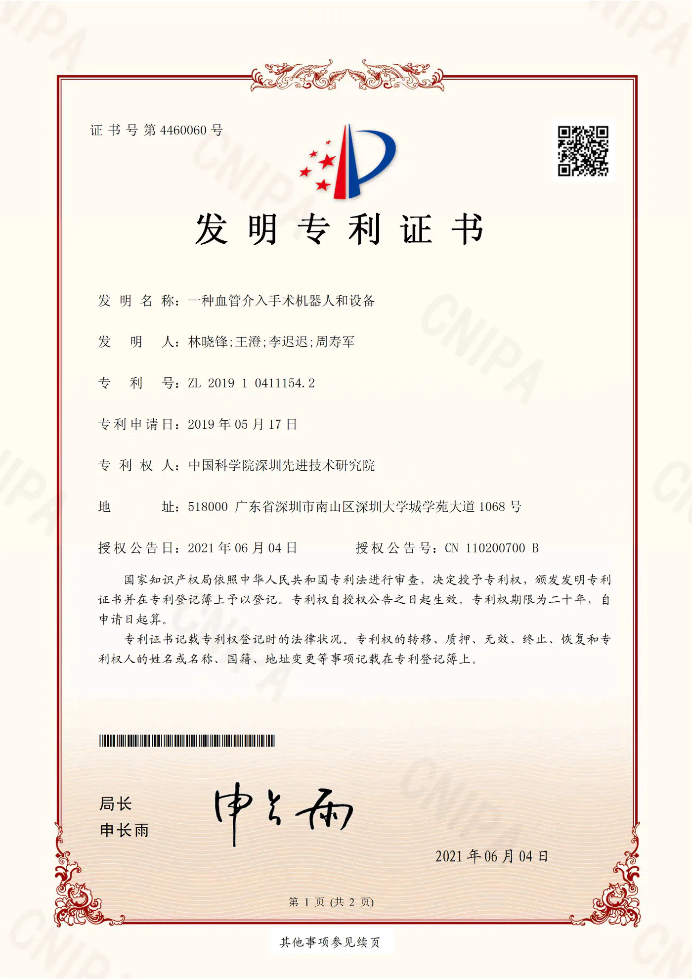
IP PORTFOLIO
Counted by patent family (the Chinese and WIPO force-feedback filings count as one). The Japanese application for a navigation system, algorithm, and instruments, together with the Chinese application for a 3D calibration board and performance-evaluation method for stereo microscopic imaging, are most closely aligned with the current product. The barrier is structural: split-path imaging × intraoperative registration × micron-scale instrument tracking, combined in one hardware-software system. The remaining patents reflect prior technical work; ownership, licensing scope, and freedom to operate require item-by-item confirmation during investment and partnership due diligence.
Team
Core members have research and academic experience at the University of Tokyo, Tsinghua University, Harvard University, Yale University, the University of Chinese Academy of Sciences, and Harbin Institute of Technology. Their disciplines span clinical medicine, computer science, and biomedical engineering, with R&D backgrounds in surgical robotics, medical imaging, and navigation systems.

FOUNDER & CEO
Researcher in medical devices, Faculty of Medicine, the University of Tokyo
 Yuhan Song
Computer Vision
Yuhan Song
Computer Vision
 Enduo Zhao
Automation
Enduo Zhao
Automation
 Shenghao Jiang
Clinical Research
Shenghao Jiang
Clinical Research
 Guanhao Lin
Tech Transfer & Fundraising
Guanhao Lin
Tech Transfer & Fundraising
 Zenghui Yu
Market & Operations
Zenghui Yu
Market & Operations
 Gina Quan
Tech Transfer & Partnerships
Gina Quan
Tech Transfer & Partnerships
 Jian Lu
Chief Advisor
Jian Lu
Chief Advisor
Computer Vision
Automation
Clinical Research
Technology Transfer & Fundraising
Market & Operations
Technology Transfer & Partnerships
CHIEF ADVISOR
Market · Competition · Current Needs
The project remains in R&D and validation. Revenue starts before approval through OEM modules and software licences, so commercialization need not wait for full-system approval; after registration the business moves to proprietary systems, software, and consumables.
01 · Position
Navigation has to keep answering two questions throughout resection: where is the instrument and what tissue is this. Existing routes each have their strengths, but all were designed for the open field; what this product does differently is to answer both questions at microscopic scale, continuously during surgery.
Soft-tissue compensation
Not microscope-ready, with compensation
Microscope-ready, with compensation
Not microscope-ready, without compensation
Microscope-ready, without compensation
Microscope-field fit →
Select any route in the chart to compare it line by line with this product; on a phone, swipe left or right to switch.
TrackingContinuous; what is tracked is an array of reflective spheres about 11 mm across, from which the tip position is derived.
RegistrationRegistered once preoperatively against a bone-fixed reference frame; after tissue shifts, registration must be repeated to update.
Microscopic fieldDoes not register or track at microscopic scale. Reflective spheres are sized for the open field, an order of magnitude away from 0.3 mm tips and a field a few millimeters wide.
HardwareSeparate infrared camera (NDI Polaris class) + reference frame + reflective spheres.
A route established in the 1990s and still the mainstream: strong at “where is the instrument”, while “what tissue is this” holds only at the moment of registration.
TrackingInstrument tracking still uses reflective spheres.
RegistrationNo implanted markers; registration is based on one surface scan and is repeated by rescanning after tissue shifts.
Microscopic fieldDoes not register or track at microscopic scale. Surface scanning is designed for the open field; a field a few millimeters wide offers limited stable features.
HardwareStructured-light projector + camera, with instrument tracking added separately.
It removes implanted markers, but each registration is still a static step rather than continuous updating as tissue deforms.
TrackingContinuous.
RegistrationRepeatable intraoperative registration with soft-tissue deformation compensation.
Microscopic fieldDoes not register or track at microscopic scale. Designed for the open field; its published indication is spine.
HardwareMulti-camera intraoperative imaging array + instrument tracking, two optical systems.
The closest approach to this product and the benchmark we take most seriously: intraoperative registration, deformation compensation, and tracking are all present. The difference is that it addresses the open field, while this product brings all three into the single optical path of the microscope.
TrackingNo instrument tracking.
RegistrationNo registration to preoperative imaging.
Microscopic fieldWorks within the microscopic field. It is itself a digital operating microscope, positioned as visualization enhancement.
HardwareDigital microscope imaging + head-mounted or screen display.
It addresses “seeing more clearly”, a different product category from the two questions navigation must answer; it sits outside this divide.
Criteria: “microscope-field fit” means whether intraoperative registration and instrument tracking are performed within a field a few millimeters wide at sub-millimeter accuracy; “soft-tissue compensation” means whether registration updates automatically after tissue shifts. Both axes are read straight off the two columns of the same name in the comparison table; no other criteria are introduced. Per-product values and sources are in the table below.
| Product / Country | Product | Technical route | Registration method | Microscopic-field fit | Soft-tissue compensation | Closed-loop capability | Standard indications |
|---|---|---|---|---|---|---|---|
| Proprio Paradigm (United States) |  | Multi-camera intraoperative imaging + tracking | Bone-fixed | Not compatible | Yes | Registration + tracking | Spine |
| Brainlab Microscope Navigation (Germany) |  | NDI-class infrared optical tracking | Bone-fixed | Not compatible | No | Registration + tracking | General orthopedics or spine |
| Medtronic StealthStation S8 (United States / Ireland) |  | NDI-class infrared optical tracking | Bone-fixed | Not compatible | No | Registration + tracking | General neurosurgery / general orthopedics |
| Beyeonics One (Israel, operating microscope) | 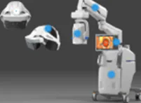 | Digital visualization, no registration | No registration | Compatible | No | Visualization enhancement only | General ophthalmology |
| Huake Precision (China) | 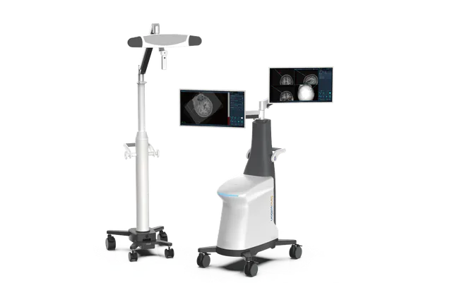 | NDI-class infrared optical tracking | Bone-fixed | Not compatible | No | Registration + tracking | General neurosurgery / general orthopedics |
| SeaSpine / Orthofix 7D FLASH (United States) |  | Structured-light registration + optical tracking | Non-invasive fixation | Not compatible | No | Registration + tracking | Neurosurgery (brain tumors) |
| Spectra Navigation Intelligent Microsurgical Navigation System (This product)Meets all three of microscopic-field fit, deformation compensation, and instrument tracking within this table |  | Coaxial beam-split dual camera | Non-invasive fixation | Compatible | Yes | Registration + tracking | General neurosurgery / ENT / ophthalmology / reconstructive surgery |
Compiled in September 2026 from each manufacturer’s public materials. Competitor specifications and approval status change over time; current specifications, indications, and registration status are subject to each manufacturer’s own disclosures. Product names and trademarks belong to their respective owners and are used here for identification only. Globally, the closest approach is Proprio Paradigm; in China, Huake Precision also targets neurosurgery but uses NDI-class infrared optical tracking, a different technical route from this product.
02 · Market → 03 · Revenue
Neurovascular, malignant brain tumor related, ENT, and non-cataract ophthalmic procedures combined.
Boundary judgment directly determines the extent of resection and survival benefit, the clearest value proposition.
Then ENT, ophthalmology, and reconstructive surgery.
Hardware module delivery; software licences bundled with hardware and billed annually; engineering services for microscope OEMs only, one-off per model. Serving university laboratories, microscope manufacturers, and navigation-system vendors to validate real demand, interface specifications, and willingness to pay.
Complete-system installations build scale; enhanced software licences renew annually; dedicated consumables launch after approval, forming recurring revenue.
04 · Risks and Current Needs
The next priority is converting technical performance into clinical evidence and a registrable product. Clinical workflow, funding, procurement logic, and the regulatory path must converge in parallel; delay in any one area will affect productization. All three risks have mitigations already under way, so none rests on a single assumption.
RESOURCE PRIORITIES
NEXT MILESTONES
The basis and sources for procedure volumes are given in the Clinical Problem section above. Market size and commercial path illustrate the commercial logic; actual performance will depend on installation ramp, gross margin, channel discounts, and the post-registration launch schedule. Each deliverable maps to a defined milestone; specific pricing, installation plans, the financing requirement, detailed use of funds, and schedule are calculated against the milestone budget, cash flow, and registration plan, and are not disclosed on this page. They are available in formal due-diligence materials.
CONTACT
We welcome direct contact from neurosurgical and microsurgical teams, microscope and instrument manufacturers, and investors following early-stage medical device programs.
Full due-diligence materials — covering the clinical validation plan, registration pathway detail, financial projections, and use of funds — are available on request. Please state your organization and intended use.
Enable JavaScript to view the email address WeChat · scan or long-press the code
SPECTRA NAVIGATION
Spectra Navigation aims to bring navigation into smaller, deeper, and more dynamic microscopic fields. The next stage requires clinical, industry, and investment partners to advance validation, registration, and scalable deployment.